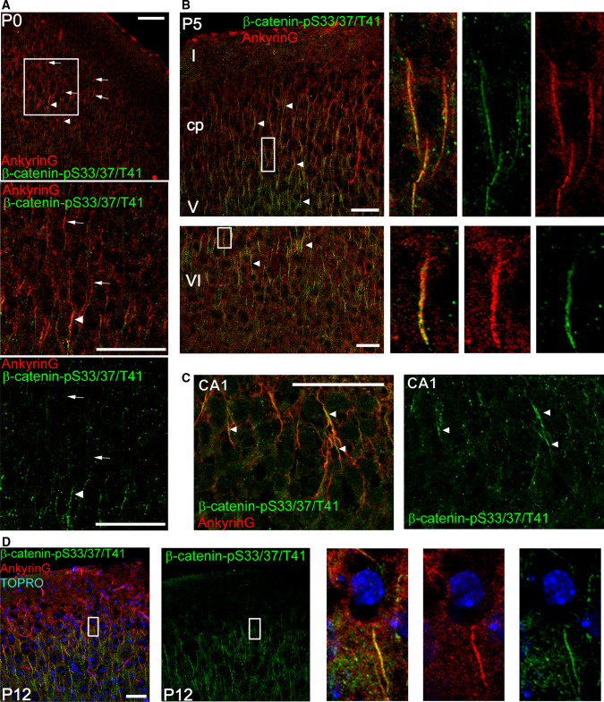
Fig. 2.
β-catenin-pS33/37/T41 is enriched at the axon initial segment in vivo. a, b Photomicrographs obtained from sections showing cortical and subcortical layers from P0 (a) and P5 (b) mice brains. c Photomicrographs of the CA1 region of a P5 mouse hippocampus. Sections were stained with anti-ankyrinG (red) and anti-β-catenin-pS33/37/T41 (green) antibodies. d Image of cortical section from P12 mouse showing β-catenin-pS33/37/T41 and ankyrinG colocalization at the AIS. Nuclei are stained with TOPRO stain. Arrows indicate ankyrinG immunolabeled AISs and arrowheads overlapping staining of β-catenin-pS33/37/T41 and ankyrinG at the AIS. Scale bar 50 µm

