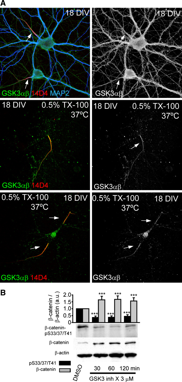
Fig. 5.
β-catenin kinase, GSK3, is tethered to the AIS after to detergent extraction. a GSK3α/β staining at the AIS of 18-DIV hippocampal neurons after detergent extraction treatment with 0.5 % Triton X-100 for 10 min (bottom panels). Upper panels show ubiquitous GSK3α/β staining in non-extracted neurons. Extracted and non-extracted neurons were stained with GSK3α/β (green), 14D4 (red) and MAP2 antibodies (blue). Arrows indicate the AIS position. b Western-blot analysis showing β-catenin and β-catenin-pS33/37/T41 total expression in 2-DIV high-density cortical neurons cultures treated with 3 µM GSK3 inhibitor X for 30, 60, and 120 min. Graph represents the mean and SEM of protein expression levels normalized to β-actin in three different experiments. ***p < 0.001, t test

