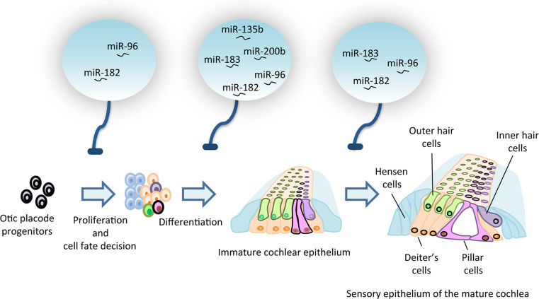
Fig. 1.
Role of miRNAs in inner ear development. The drawing depicts the development of the organ of Corti viewed in the transversal plane, showing the relative position of the inner hair cells (violet) and the three rows of sensory outer hair cells (green). The pale blue circles on the top indicate the main miRNAs implicated in the control of the different steps of inner ear development and its homeostatic role in the mature organ (see text for further details)

