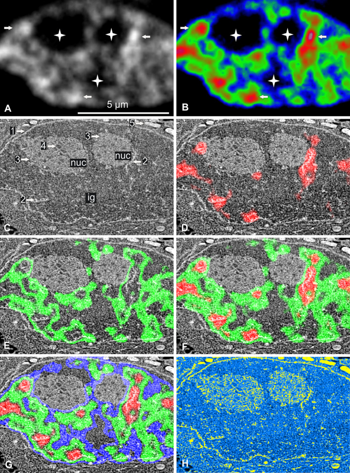
Fig. 1.
Correlative cryo-analytical scanning transmission electron microscopy of a control HeLa H2B-GFP interphase cell. a, b Fluorescence mode: a fluorescence intensity imaged as gray-scale levels or b coded in red, green, and blue for high, medium, and low fluorescence intensity, respectively. Chromatin is readily detectable within this 85-nm ultrathin cryo-section. Higher fluorescence intensities identify both rounded and elongated foci of condensed chromatin (arrows) surrounded by large irregular areas of uncondensed chromatin. Several broad areas displayed no fluorescence (stars). c Dark-field STEM image of the same freeze-dried cell. The nuclear envelope (arrow 1), several high-contrast narrow elongated structures (arrow 2), large roundish nuclear domains (arrow 3) containing tiny roundish structures with low levels of contrast (arrow 4) and mitochondria (arrow 5) can be seen. d–g Merging of the STEM image with the fluorescence intensity image: d high (red); e medium (green); f high and medium (red and green); g high, medium, and low intensity (red, green, blue). The fluorescence and STEM images were perfectly aligned, demonstrating an absence of differential shrinkage during freeze-drying. This makes it possible to identify clumps of condensed chromatin (arrow 2). The areas with no fluorescence correspond to two nucleoli (3, nuc) (containing fibrillar centers (arrow 4)) and interchromatin granules (ig). h Parametric water content image computed from the STEM image. Values in % are coded in yellow (0–50 %) and on a linear gradient ranging from light to dark blue (51–100 %). Mitochondria were the least hydrated organelles of the cell. In the nucleus, condensed chromatin (previously identified by fluorescence) was found in both narrow clumps and filaments, which, together with part of the nucleolus, were the least hydrated domains of the nucleus. Nucleolar fibrillar centers and some domains of the nucleoplasm contain the largest amounts of water. Scale bar 5 μm

