Fig. 5.
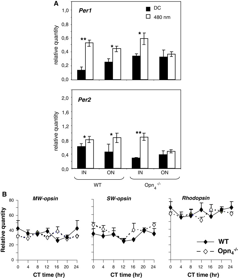
A Light induction of Per1 and Per2 in the retina of wild-type and Opn −/−4 mice using a pulse of monochromatic light (480 nm, 15 min, 2.8 × 10¹⁴ photons/cm2/s) administered at CT16. Animals were sacrificed 30 min after the beginning of the light pulse. The inner (IN) and the outer (ON) regions of the retina were isolated by laser microdissection and mRNA levels were measured using real-time reverse transcription-polymerase chain reaction. Results are expressed as mean ± SEM (n = 5 for each genotype). Asterisks indicate a statistically significant difference (ANOVA: p < 0.05; post hoc Newman-Keuls tests comparing genotypes between the IN and ON retinas (*p < 0.05, **p < 0.01). B Expression of middle-wavelength (MW), short-wavelength (SW) opsins and rhodopsin mRNA in the retina of wild-type (black diamonds) and Opn −/−4 (white diamonds) mouse kept under DD cycle. Transcripts were measured by real-time reverse transcription-polymerase chain reaction, expression was normalized to HPRT control gene. For both genotype, transcripts are not rhythmic over the 24-h cycle in the retina and for each CT, no significant differences between genotype are observed (ANOVA: p > 0.05)

