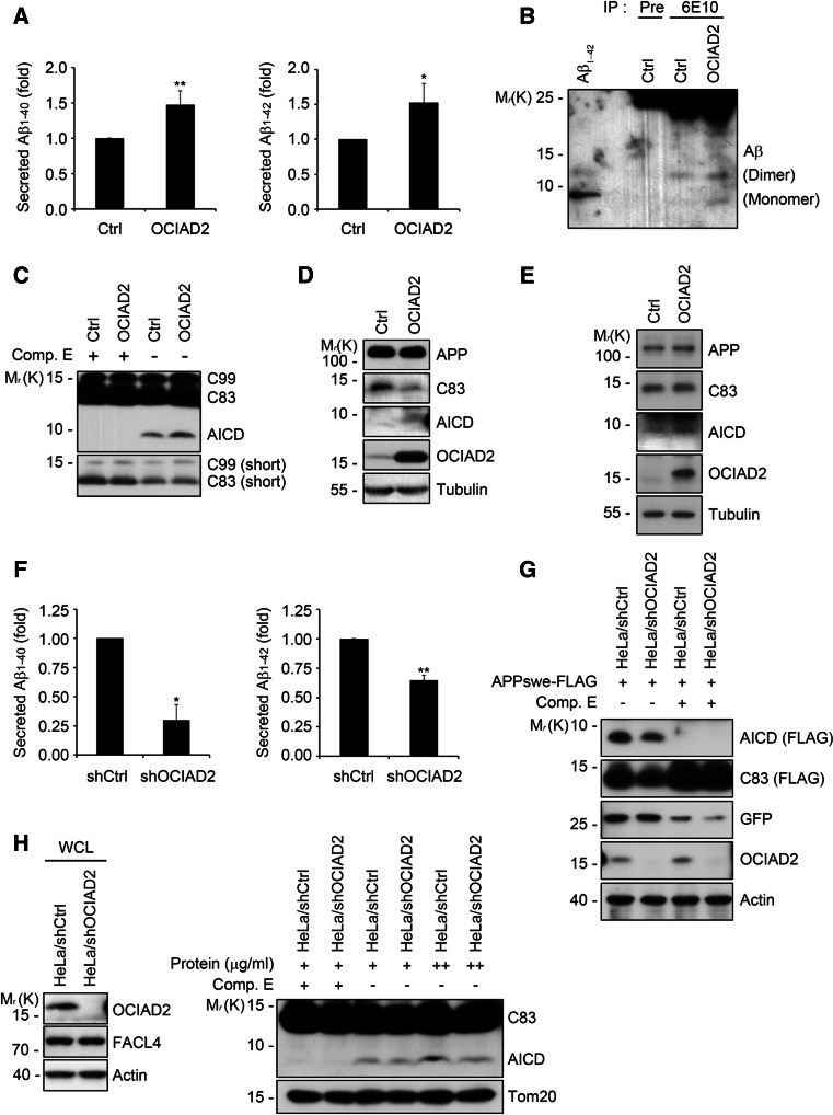
Fig. 1.
OCIAD2 increases Aβ generation. a OCIAD2 overexpression stimulates Aβ1–40 and Aβ1–42 generation in cells. SH-SY5Y-APPswe cells were transfected with pcDNA3 (pCtrl) or OCIAD2 (pOCIAD2), and maintained in the conditioned media for 48 h. The media was measured for Aβ1–40 (left) and Aβ1–42 (right) using ELISA kit. Bars mean ± SD (n = 5). *P < 0.05, **P < 0.01. b OCIAD2 overexpression increases the production of Aβ species. CHO-7PA2 cells were transfected with pCtrl or pOCIAD2 for 36 h and then incubated in serum-free DMEM for another 12 h. The media was subjected to immunoprecipitation (IP) assay using pre-immune serum (Pre) or anti-Aβ antibody (6E10), and the immunoprecipitates were analyzed with western blotting. Monomer and dimer of Aβ are indicated. c, d OCIAD2 overexpression increases the generation of AICD. HEK-APP695 cells were transfected with pCtrl or pOCIAD2 for 48 h and crude membrane fraction was prepared by centrifugation as described in “Materials and methods”. The membrane fraction (10 μg) was then incubated at 37 °C for 2 h with or without 100 μM Compound E (Comp. E) and separated by SDS-PAGE for western blotting (c). The whole cell lysates from the homogenized sample from (c) were subjected to SDS-PAGE and analyzed by western blotting (d). e Ectopic expression of OCIAD2 enhances AICD generation in BACE KO MEF cells. BACE KO MEF cells were transfected with either pCtrl or pOCIAD2 for 48 h and then subjected to western blot analysis. f OCIAD2 knockdown reduces Aβ1–40 and Aβ1–42 production. HeLa/pSuper-neo (shCtrl) and HeLa/pSuper-neo-shOCIAD2 (shOCIAD2) stable cells were transfected with pAPPswe-FLAG and pEGFP for 72 h in the conditioned media, and Aβ1–40 (left) and Aβ1–42 (right) were then measured using ELISA kit. Bars mean ± SD (n = 3). *P < 0.05, **P < 0.01. g, h OCIAD2 knockdown reduces AICD generation. After co-transfection of HeLa/shCtrl and HeLa/shOCIAD2 stable cells with pAPPswe-FLAG and pEGFP for 24 h, cell extracts were analyzed by western blotting (g). Membrane fraction (30 and 50 μg) prepared from HeLa/shCtrl and HeLa/shOCIAD2 stable cells was incubated at 37 °C for 2 h with or without 100 μM Comp. E and then analyzed by western blotting (h, right). Expression of OCIAD2 was determined by western blotting (h, left)

