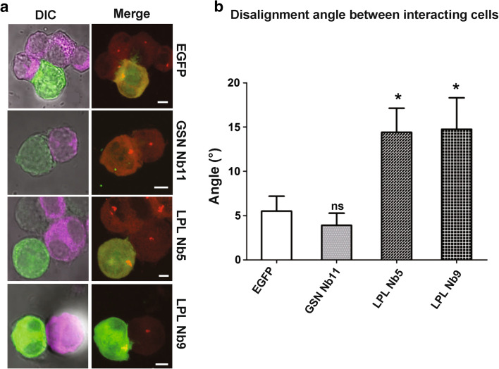
Fig. 5.
LPL Nbs promote a defect in centrosome docking. Jurkat cells transfected with EGFP, GSNNb11-, LPL Nb5- or LPL Nb9-EGFP were incubated during 45 min with SEE-pulsed Raji cells which were prelabeled with a fluorescent far red dye. a Confocal images of conjugated cells expressing LPL/GSN Nbs or EGFP. Left panel Raji cells are shown in magenta (far red) and transfected Jurkat cells in green (EGFP). Right panel Z-dimensional acquisition for gamma-tubulin (red) and EGFP (green) staining, followed by projection on the Z-axis. Bar 5 μm. b Bar graph representing the angle measurement between the X-axis and the line linking the centrosome to the center point of the cell extremity (see “Materials and Methods”). Representation of conjugated cells (n = 20) angle measurements for the different conditions from three independent experiments. Error bars mean ± SEM. Unpaired t tests were performed to observe statistical differences in the area between the cells expressing EGFP and the cells expressing (different) nanobodies (*p < 0.05; **p < 0.01; ***p < 0.001)

