
Fig. 6.
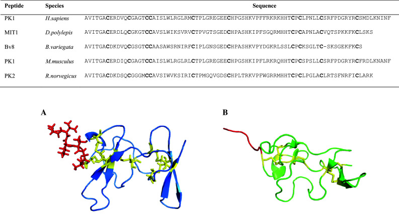
Sequence alignment and 3D structures of AVIT proteins. Cysteine residues are shown in bold. Accession numbers are as follows: P58294, P25687, Q9PW66, Q14A28, Q8R413. A NMR structure of MIT1 (1IMT) highlighting the AVITGA residues in stick format (red). B NMR structure of Bv8 from B.variegata (2KRA) showing the AVITGA N-terminal region in red. Disulfide bridges are depicted in yellow