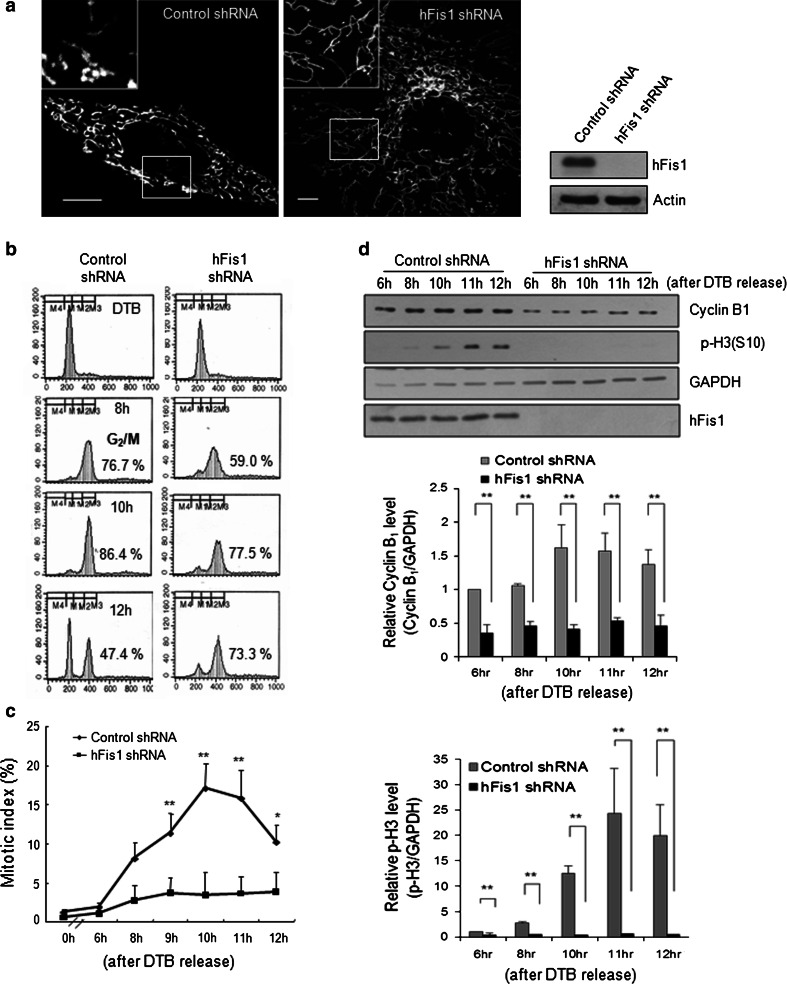
Fig. 2.
Highly elongated mitochondria by inhibition of mitochondrial fission failed to enter mitosis. HeLa cells were transfected with pREP4 construct containing short hairpin RNA (shRNA) of the target sequence of hFis1 and the transfectants were selected by growing in media containing hygromycin B. Mitochondrial morphology was analyzed by confocal microscopy. a Elongated net-like mitochondrial morphology in hFis1-depleted cells was visualized by MitoTracker staining (left) and evaluation of hFis1 knockdown by immunoblotting (right). Mitochondrial morphology was analyzed by confocal microscopy. b hFis1-depleted cells were synchronized at G1/S boundary using double thymidine block (DTB) method and released from DTB. Synchronized HeLa cell extracts were harvested at the indicated times after thymidine release and DNA contents were analyzed by flow cytometry. c Cells were harvested at the indicated time point after DTB release and stained with aceto-orcein. Mitotic index was determined by counting cells with condensed chromatin under optical microscopy. At least 200 cells were counted under each condition and repeated three times to calculate mean standard deviation. d Cell lysates for analysis of cyclin B1/cyclin-dependent kinase1 (Cdk1) kinase activity were harvested at each time point and the expression levels of cyclin B1 and phospho-histone H3 (p-H3) were analyzed by immunoblotting. Anti-cyclin B1 antibody was used to immunoprecipitate kinase complexes and histone H1 was used as a substrate. Phosphorylated substrates were detected by autoradiography. The graphs represent quantification of cyclin B1 or p-H3 expression levels by normalization to GAPDH. *p < 0.05, **p < 0.01 vs. control shRNA by Student’s t test

