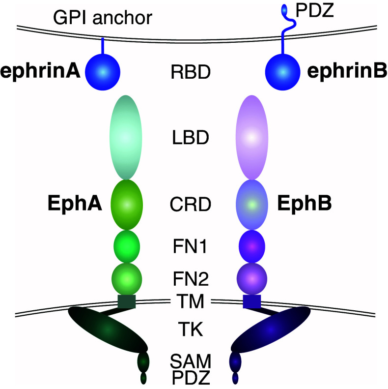
Fig. 1.
Structure of Eph receptors and ephrin ligands. EphA and EphB receptors share a conserved multi-domain structure [24]. The extracellular domain contains an N-terminal ligand-binding domain (LBD), a cysteine-rich domain (CRD) followed by two fibronectin type-III repeats (FN1 and FN2). A single-pass transmembrane domain (TM) is followed by an intracellular region containing a juxtamembrane region, a tyrosine kinase domain (TK), a sterile α motif (SAM), and a postsynaptic density protein PSD95, Drosophila disc large tumor suppressor DlgA, and zonula occludens-1 protein ZO-1 (PDZ)-binding motif [3, 6]. The ephrin ligands contain a conserved extracellular N-terminal receptor binding domain (RBD). EphrinA ligands are attached to the cell membrane through a glycosylphosphatidylinositol (GPI)-anchor, whereas ephrinBs contain a transmembrane domain, and a C-terminal cytoplasmic tail including a PDZ-binding motif [29]

