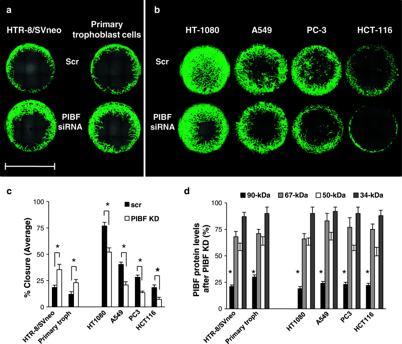
Fig. 1.

The effect of PIBF knock-down on invasiveness of trophoblast and tumor cells. The HTR-8/SVneo trophoblast cell line and primary trophoblast cells isolated from first-trimester abortion material (a) as well as HT-1080 (fibrosarcoma), A549 (lung carcinoma), HCT-116 (colorectal cancer), PC-3 (prostate cancer) tumor cell lines (b) were transfected with PIBF siRNA, or scrambled oligonucleotides (scr). Wells were seeded with control (scr) or PIBF siRNA-treated cells. Invasion of cells into the detection zones after 72 h is shown. Cells were stained with calcein AM. Images were captured using multiarea scan by a confocal microscope (bar 2 mm). c Quantification of area closure (%) calculated from measured areas of cell invasion at 72 h. Data are presented as mean ± SEM from 12 wells (HTR-8/SVneo, HT-1080) or six wells (other cell lines and primary trophoblast cells) per condition (asterisk indicates significant difference from scrambled at p < 0.05). d The efficiency of PIBF knock down (KD) was controlled by measuring PIBF levels (90-, 67-, 50-, and 34-kDa isoforms) using Western blotting at each individual experiment. Statistical analysis of Western blots are shown as mean ± SEM; asterisk indicates significant difference from scrambled at p < 0.05
