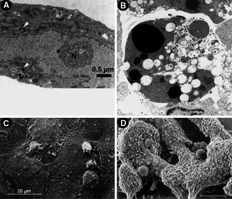
Fig. 1.

Transmission (a, b) and scanning electron micrographs (c, d) of Stx-challenged human endothelial cells. a Uptake of gold-labeled Stx2 in HUVECs after 4 h of toxin treatment (taken from [109]). Black beads marked intracellularly with white arrowheads, correspond to gold-labeled Stx2. b Morphological features of human dermal (foreskin) microvascular endothelial cells after 16 h exposure to Stx. Magnification ×7,500 (taken from [138]). c Leukocyte adhesion and transmigration in HUVECs under flow after challenge for 24 h with Stx2 (drawn from [129]). d Formation of organized thrombi with entrapped leukocytes on human microvascular endothelial cells after incubation for 24 h with Stx1 (drawn from [116]). Compilation of four original figures, reprinted with permission from Matussek et al. [109], copyright The American Society of Hematology 2003 (a), Pijpers et al. [138], copyright The American Society of Nephrology 2001 (b), adapted by permission from Macmillan Publishers (Kidney International) Zoja et al. [129], copyright The International Society of Nephrology 2002 (c), and Morigi et al. [116], copyright The American Society of Hematology 2001 (d)
