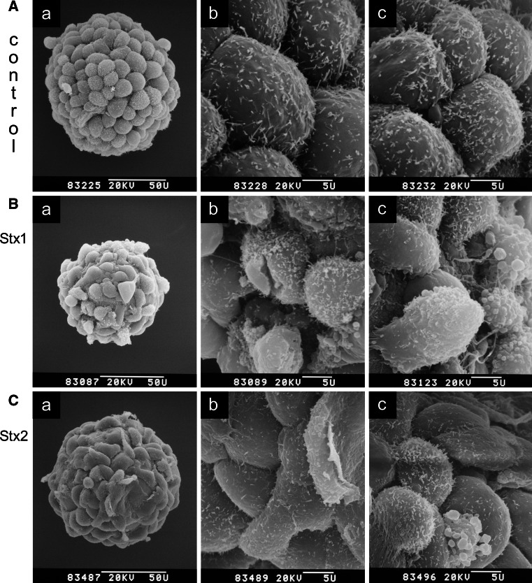
Fig. 2.
Scanning electron microscopy of Stx1- and Stx2-treated HBMECs. Cells were grown on collagen-coated microcarriers and confluent monolayers (a a–d, controls) were exposed for 48 h to 500 ng/ml of Stx1 (b a–c) or 500 ng/ml of Stx2 (c a–c). The a panels show the microcarrier overview screens and the b panels examples of corresponding higher magnified partial views of the same microcarriers. The c panels of the a–c series show partial views of microcarriers from parallel cell cultures incubated under identical conditions. Bars 50 μm (50 U) or 5 μm (5 U) as indicated in the micrographs. Original electron-optical magnifications of the microcarrier overview screens are ×870 (a a), ×970 (b a), and ×980 (c a). Magnifications of partial detail views are ×4,500 of a–c. Compilation of an original figure, reprinted with permission from Bauwens et al. [146], copyright 2010 Schattauer

