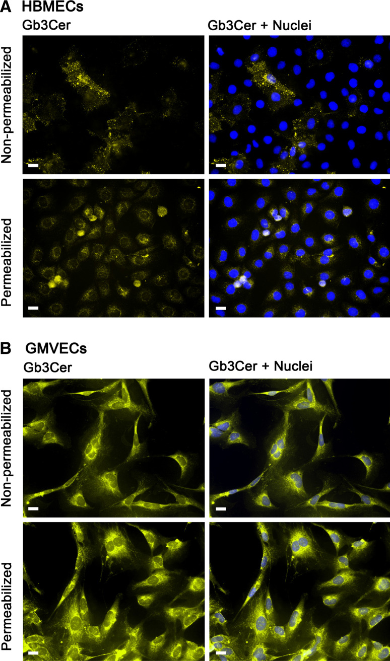
Fig. 8.
Indirect immunofluorescence microscopic detection of Gb3Cer in HBMECs and GMVECs. Subconfluent grown HBMECs (a) and GMVECs (b) were immunostained with monoclonal rat 38.13 anti-Gb3Cer antibody followed by incubation with Alexa Fluor® 546 labeled secondary antibody after permeabilization with 0.3 % Triton X-100 (lower panels) and as non-permeabilized cells (upper panels). Cell nuclei were stained with 4′,6-diamidino-2-phenylindol (DAPI). Bars 20 μm

