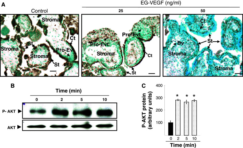
Fig. 4.

EG-VEGF effect on placental villi survival. a Representative photographs of TUNEL staining (in brown) in PEX that have been treated or not with EG-VEGF. PEX (10 wg) were serum starved for 72 h and treated for 24 h with EG-VEGF (25 and 50 ng/ml). EG-VEGF condition shows much less TUNEL-positive cells compared to the control condition. b Representative Western blot of AKT phosphorylation after treatment with 50 ng/ml EG-VEGF. Standardization of the protein signals was done with antibodies against total-AKT. Quantification of the intensity of the bands is illustrated in c. Data represent the mean ± SEM of triplicates *p < 0.05 vs. control. Scale bar: 20 μm. Ct cytotrophoblast, St syncytiotrophoblast, Bv blood vessel, Pro-Evt proliferative extravillous trophoblasts
