Fig. 1.
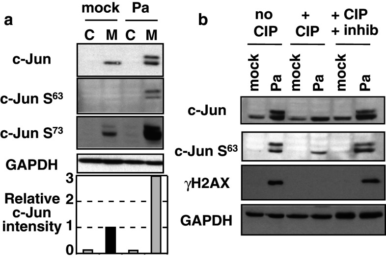
Hyperphosphorylation of c-Jun in HL60 macrophages in response to P. aeruginosa. a Undifferentiated control HL60 cells (C) or HL60 macrophages (M) were infected (or not, mock) with P. aeruginosa CHA strain (Pa) at a MOI of 10. Two and half hours post-infection, cell extracts were prepared and analyzed by Western blot with the indicated antibodies. Blots were quantified with ImageJ and c-Jun relative intensity normalized to GAPDH level was set to 1 (dark bar) in mock macrophages. b Cell extracts from HL60 macrophages infected as above (Pa) or not infected (mock) were incubated with CIP or with CIP in the presence of phosphatase inhibitors (CIP + Inhib) and then analyzed by Western blot

