
Fig. 2.
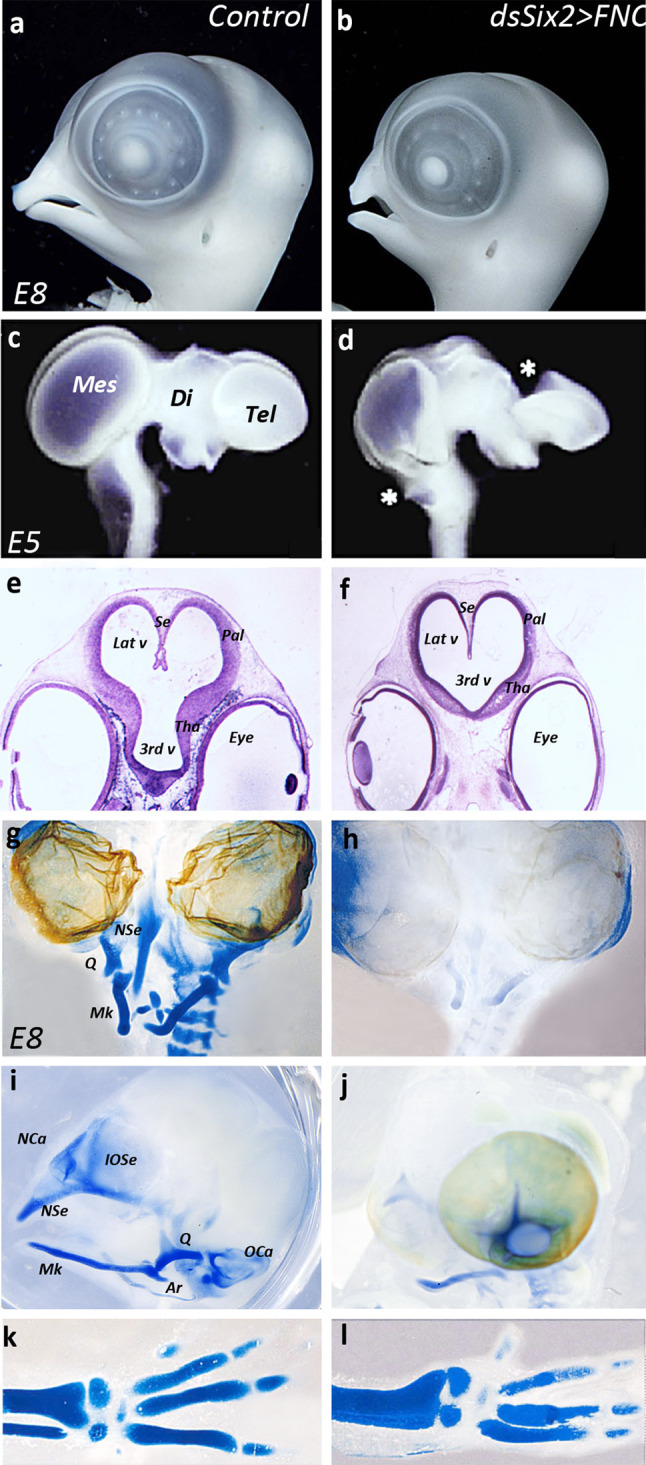
Six2-silencing in the FNC hampers head and brain development. a, b Morphology and c, d whole-mount brain preparation in control (a, c) and experimental (b, d) embryos. In Six2-silenced embryos, the nasofrontal and maxillo-mandiblar development is atrophied (b), and brain morphology is altered along the dorsal midline (d): choroid plexi are missing (stars). Cresyl-violet staining of paraffin sections (e, f) on E5 control (e) and experimental (f) embryos showing the defective development of prosencephalic structures in experimental series: the pallial and thalamic neuroepithelium are thinner than in control (f). In addition, the inter-hemispheric septum fails to form normally. g–j Whole-mount staining of cartilage in the head skeleton of E8 (g, i) control and (h, j) Six2-silenced embryos. On frontal views (g, h), much of the nasal and mandibular cartilages are absent in experimental embryos (h). On profile views (i, j), the skeletal defects resulting from the silencing of Six2 consist in the absence of the interorbitary and nasal septa, nasal and otic capsules, quadrate and articular (j); on control embryo, eyeballs have been removed to evidence skeletal structures at the midline. By contrast, the development of the appendicular skeleton is similar in the control and Six2-silenced series, as shown by the normal development of the radius, ulna, and digits (k, l). Ar articular, Di diencephalon, IOSe interorbital septum, Lat v lateral ventricle, Mes mesencephalon, Mk Meckel’s cartilage, NCa nasal capsule, NSe nasal septum, OCa otic capsule, Pal pallium, Q quadrate, Se septum, Tel telencephalon, Tha thalamus, 3rd v third ventricule