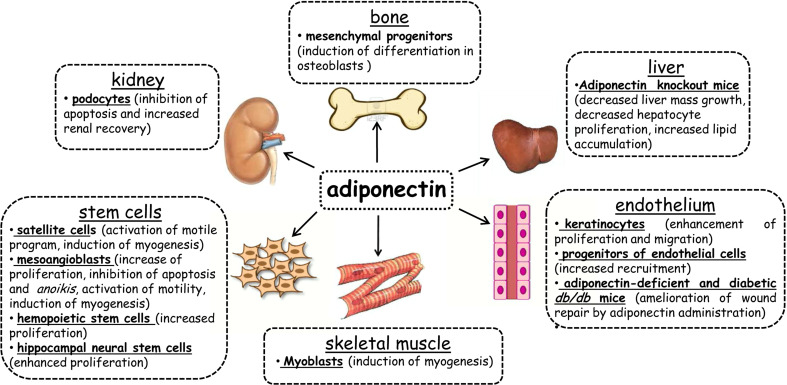
Fig. 2.
Adiponectin participates in tissue regeneration. The figure shows the cell lines and/or the animal models used to demonstrate the involvement of adiponectin in tissue regeneration. The obtained results for each tissue are reported (detailed text in paragraph “Role of adiponectin in the regeneration of non-muscle tissues”). Both fAd and gAd play a role in tissue regeneration. gAd acts on satellite cells in skeletal muscle by inducing cell motility and myogenesis [15]; on mesoangioblasts by activating proliferation, cell motility, myogenesis, and inhibiting both apoptosis and anoikis [60]; on hemopoietic stem cells [71] and hippocampal neural stem cells by inducing cell proliferation [72]. In skeletal muscle, gAd promotes the differentiation of myoblasts into myotubes [40]. In the kidney, fAd acts by inhibiting apoptosis in podocytes and by supporting renal recovery [76]. Depletion of fAd in murine liver leads to a decrease in mass growth and hepatocyte proliferation [74] and an increase in lipid accumulation [75]. In the bone, fAd promotes the differentiation of mesenchymal progenitors into osteoblasts [77]. In the endothelium, fAd promotes the enhancement of proliferation and the migration of keratinocytes [88], ameliorates wound repair in adiponectin-deficient and diabetic db/db mice [88] and increases the recruitment of endothelial cell precursors [78]

