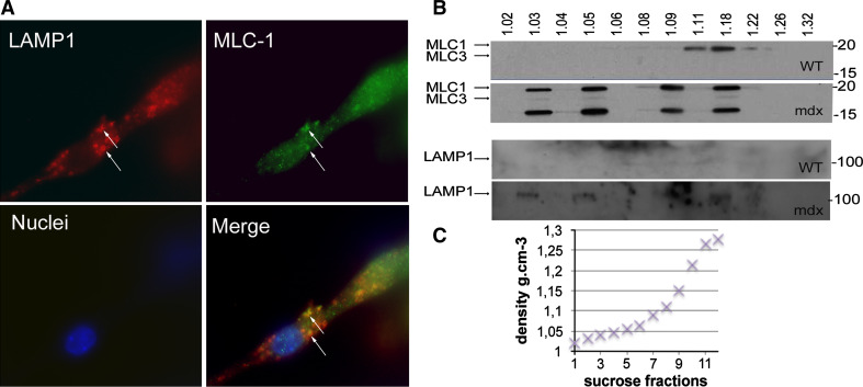
Fig. 6.
Colocalization of LAMP1 and MLC1 in mdx myotubes. a BacMam system (Invitrogen) was used to deliver a cDNA encoding fluorescently tagged LAMP1, and MLC1 was immunolabeled. MLC1 is in green, LAMP1 in red, and nuclei in blue (×63 objective). Arrows colocalization of LAMP1 and MLC1. WT wild-type, MLC1 myosin light chain 1. b Immunoblots of MLC1-3 (upper blots) and of LAMP1 (lower blots) on vesicle fractions extracted from the culture media of wild-type and mdx myotubes. Vesicles were isolated using sucrose gradient suspension under ultracentrifugation. The sucrose density (g cm−3) of the 12 fractions is indicated above the Western blot. The lower blot shows an aberrant distribution of vesicle densities in mdx culture medium. The molecular weight (kDa) is indicated on the right of each blot. c Plot of fraction density against fraction number for sucrose gradient

