Fig. 4.
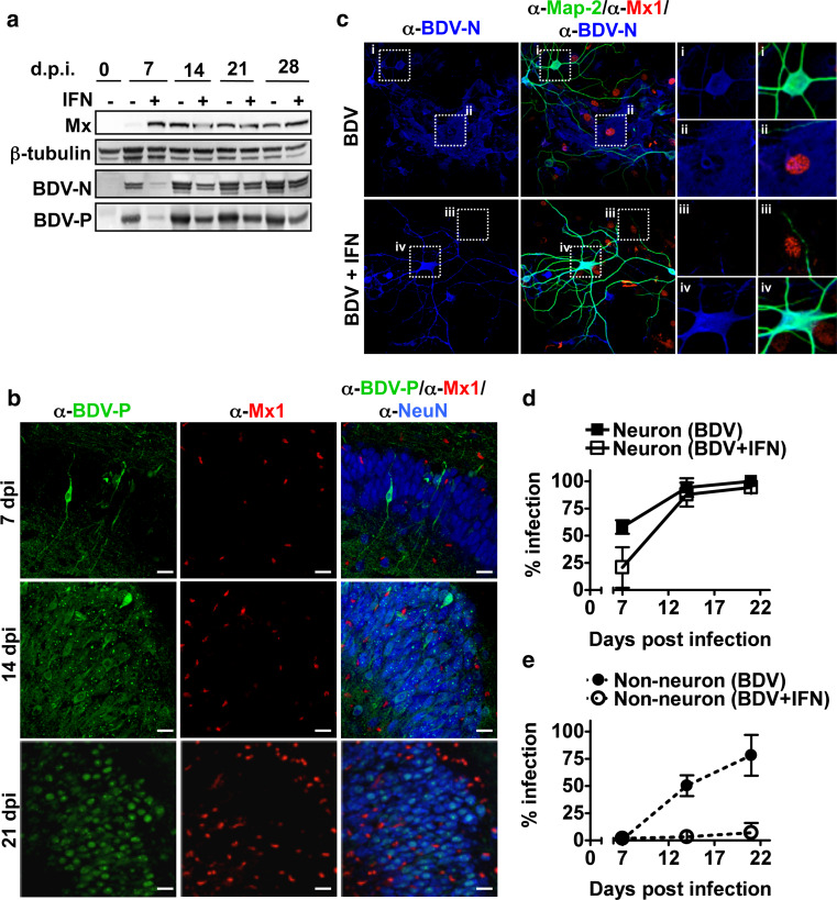
BDV replication in neurons is not abrogated by treatment with type I IFN. a BDV-infected hippocampal slice cultures from SD rats were either treated or untreated with IFN-α (105 U/ml) after 3 days of virus infection. The medium was changed three times per week over 3 weeks. At the indicated time point post-infection, the cell extract was prepared from a pool of at least four cultures and analyzed by Western blotting for the presence of the indicated proteins. b Staining of hippocampal sections of BDV-infected rat slice cultures at the indicated time point p.i. using antiserum against BDV-P together with antibodies against Mx1 (α-Mx1) and NeuN (α-NeuN), a neuron-specific marker. Scale bars 20 μm. Upper panel CA1 pyramidal layer; Middle and lower panel CA3 pyramidal layer. c Dissociated neuronal cultures from the hippocampus of SD rats were infected with BDV (1,000 FFU) in the presence or absence of IFN-α for 21 days and stained for the presence of BDV-N (α-BDV-N), Mx1 (α-Mx1) and a neuronal marker Map-2 (α-Map-2). Scale bars 30 μm. Right four panels (i–iv) represent magnifications of the indicated areas. The infection rate of neurons (d) or non-neuronal cells (e) was determined by counting BDV-positive cells in Map-2-positive and -negative cells, respectively

