Fig. 2.

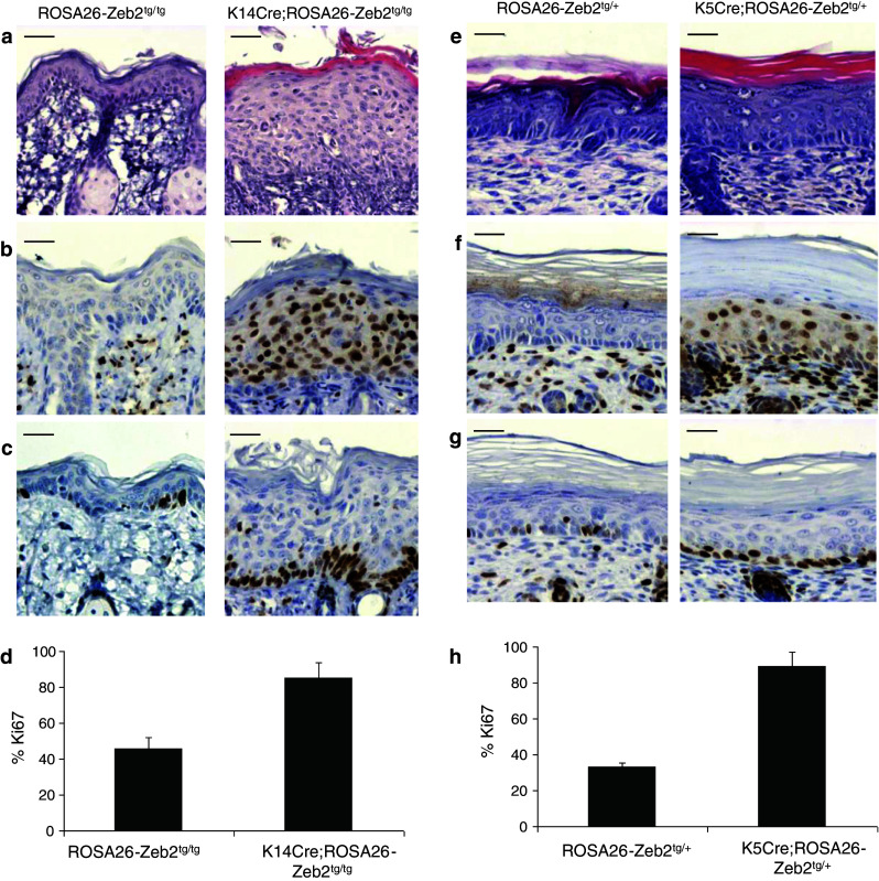
Expression of Zeb2 in the epidermis of K14Cre;ROSA26-Zeb2 tg/tg and K5Cre;ROSA26-Zeb2 tg/+mice. Hematoxylin-eosin staining on wild-type and transgenic skin revealed that Zeb2 expression in the epidermis results in hyperproliferation in adult K14Cre;ROSA26-Zeb2 tg/tg mice (a) and in K5Cre;ROSA26-Zeb2 tg/+ P4.5 neonates (e). Zeb2 is present in all epidermal layers in both strains (b and f) as seen by immunohistochemistry using a mouse Zeb2-specific antibody. Analysis for the proliferation marker Ki67 revealed that the basal layer is actively proliferating in both strains (c and g). Quantification of the number of Ki67-positive cells confirmed this observation (d and h). The graphs depict the average percentage ± SD of Ki67-positive cells in the basal layer (100 %) counted in four different low magnification photographs (p < 0.001 in both d and h). Bars 40 μm
