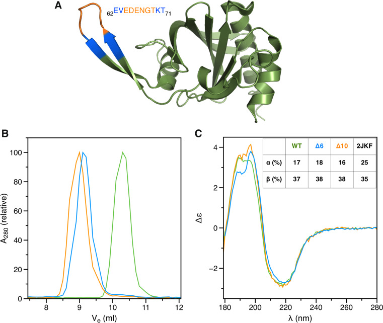
Fig. 1.
Dimerization of profilin mutants. a Structure of the wild-type P. falciparum profilin (2JKF; [29]). Residues deleted in the different mutants are indicated with colors: orange Δ6; blue Δ10. b Size-exclusion chromatogram showing dimerization of the deletion mutants (green wild-type profilin; orange Δ6; blue Δ10). c SRCD spectra of the wild-type and mutant profilins (colors as above). Secondary structure contents calculated from the spectra using DichroWeb and as in the wild-type crystal structure (2JKF) are shown in the inset

