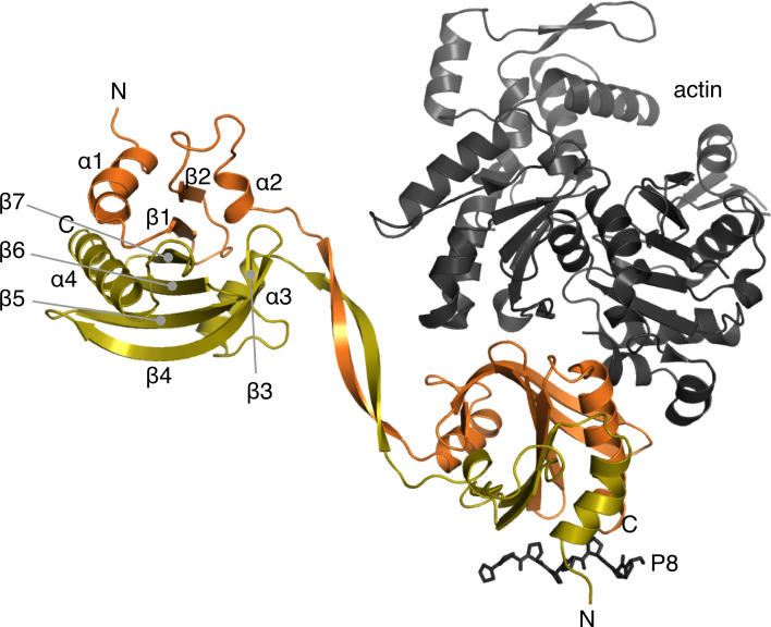
Fig. 3.
Dimerization mode of the domain-swapped Δ6 mutant. The two chains are colored orange and yellow. The secondary structure elements of the core profilin fold are labeled in the upper left subunit. The N and C termini are labeled in both subunits. Actin and an octa-proline peptide are depicted on the lower right subunit as they are bound to a canonical profilin (PDB code: 2PAV [76]) and the wild-type P. falciparum profilin (PDB code: 2JKG [29]), respectively

