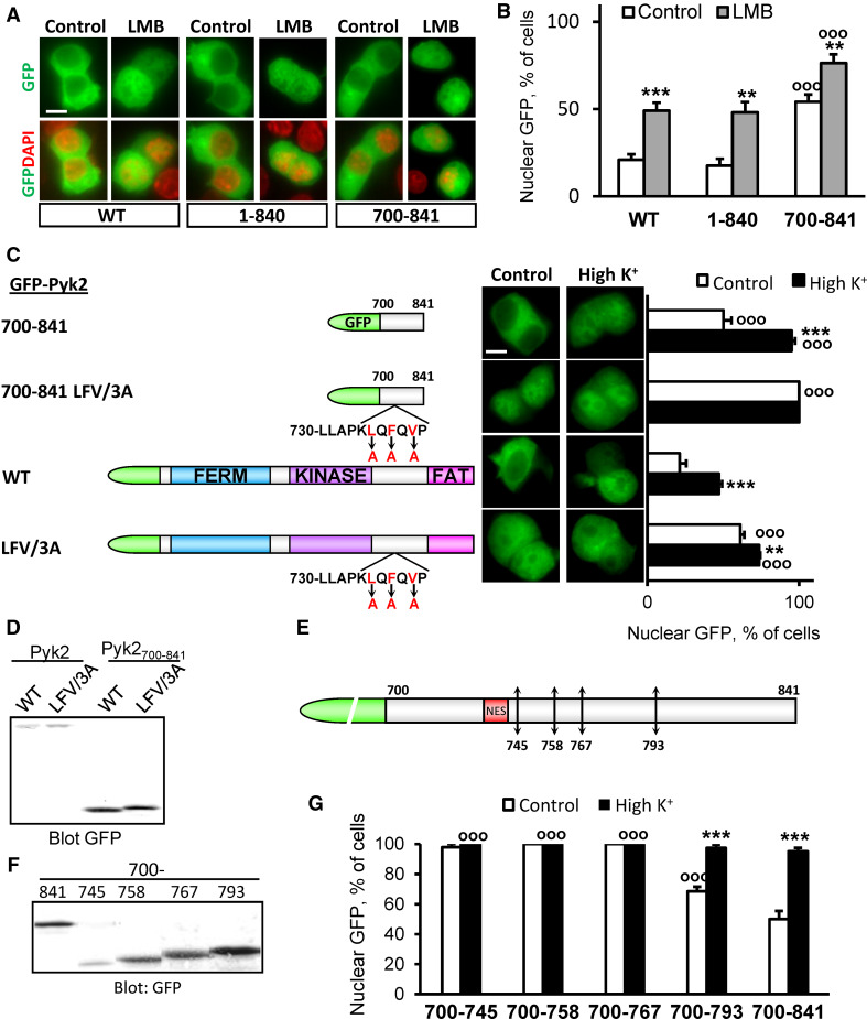
Fig. 3.
Pyk2700–841 contains a nuclear export motif. a PC12 cells transfected with GFP-Pyk2 (WT), 1–840 or 700–841 were incubated in the absence or presence of LMB (11 ng/ml, 3 h). GFP-Pyk2 fluorescence and nuclei stained with DAPI were analyzed. b Percentage of cells with nuclear GFP. Values are means + SEM, two-way ANOVA: deletion effect F (2,20) = 31.09, p < 0.0001, LMB effect F (1,20) = 48.88, p < 0.0001, interaction F (2,20) = 0.39, p > 0.05. Newman–Keuls test: **p < 0.01, ***p < 0.001 versus control. °°°p < 0.001 compared to WT. c PC12 cells transfected with WT or LFV/3A GFP-Pyk2 or GFP-Pyk2700–841 (scheme on the left) were treated with high K+ (High K+) or control solution for 3 min. GFP fluorescence and nuclei stained with DAPI were analyzed (middle) and the percentage of cells with n ≥ c GFP quantified (right). Values are means + SEM, two-way ANOVA: deletion effect F (4,30) = 158.77, p < 0.0001, depolarization effect F (1,30) = 113.22, p < 0.0001, interaction F (4,30) = 21.71, p < 0.0001. Newman–Keuls test: **p < 0.01, ***p < 0.001 versus control. °°°p < 0.001 versus WT. d Immunoblot analysis of constructs as in c with anti-GFP or anti-Pyk2 antibody. e Positions of stop codons inserted in GFP-Pyk2700–841 (745, 758, 767 and 793). f PC12 cells transfected with the constructs presented in e were analyzed by immunoblotting with anti-GFP. g Percentage of transfected cells with n ≥ c GFP. Values are means + SEM, two-way ANOVA: deletions effect F (4,25) = 47.88, p < 0.0001, depolarization effect F (1,25) = 82.58, p < 0.0001, interaction F (4,25) = 32.49, p < 0.0001. Newman–Keuls test: ***p < 0.001 versus control. °°°p < 0.001 versus 700–841. Scale bar 5 μm

