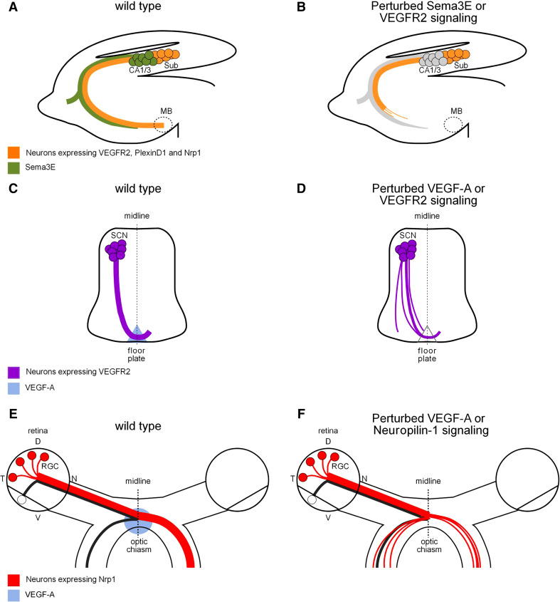
Fig. 3.
Novel role for VEGF-A in axonal wiring. a Schematic representation of the pathways taken by subiculo-mammillary projections in the developing mouse brain. VEGFR2 expression by subicular axons is involved in the recognition mechanism of the attractive/growth-promoting factor Sema3E, supplied locally by efferent CA1/3 axons. b Genetic ablation of Sema3E or neural VEGFR2 results in a hypoplastic subiculo-mammillary tract with few axons reaching their appropriate target, even at adult stages. c Schematic of spinal commissural axon projection toward and across the floor plate in the mouse embryo. Commissural axons express VEGFR2 and are attracted to the ventral midline by VEGF-A secreted from the floor plate. d Deleting VEGFR2 in spinal commissural neurons or lowering VEGF-A levels in the floor plate cause commissural axon pathway defects, including defasciculation and axonal misprojections to the lateral edge of the spinal cord. e Schematic representation of the routing of retinal ganglion cell axons at the optic chiasm to the appropriate hemisphere of the mouse brain. Ganglion cells giving rise to uncrossed axons are located in the ventrotemporal retina, whereas ganglion cells in the other retinal quadrants cross over the optic chiasm. Crossing axons express Neuropilin-1 and are guided across the optic chiasm by VEGF-A164. f Disrupted VEGF-A164-Neuropilin-1 signaling induces axon defasciculation and ipsilateral misprojections. CA1/3 Cornu Ammonis 1 and 3; D dorsal retina; MB mammillary bodies; N nasal retina; RGC retinal ganglion cells; SCN spinal commissural neurons; Sub subiculum; T temporal retina; V ventral retina

