Fig. 4.
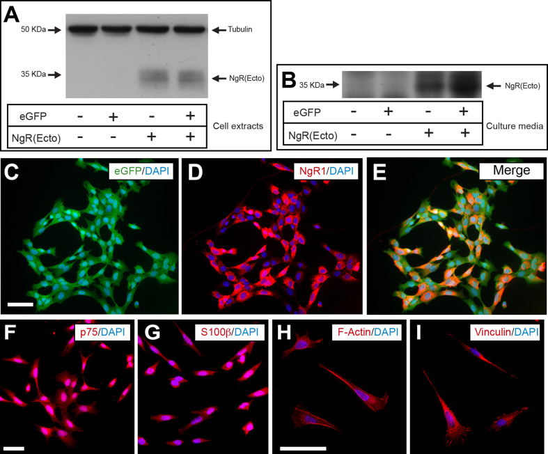
Generation of the TEG3 cell lines overexpressing NgR(Ecto). a Western blot illustrating the expression of the NgR(Ecto) in TEG3 cells after lentiviral delivery of eGFP (+ or −) and NgR(Ecto) (+ or −). Arrows point to the proteins of interest. Tubulin was immunoblotted as control protein. b Western blotting illustrating the presence of NgR(Ecto) in the culture media of TEG3 cells after the lentiviral delivery of eGFP and NgR(Ecto) as above. c–e Example of the expression of eGFP (e) and NgR1 (i) in double-infected cells. f–i Fluorescence photomicrographs of eGFP-NgR(Ecto)-TEG3 cells immunoreacted to p75 (f), S100β (g), F-Actin (h), and vinculin (i) detection. Scale bars c and f = 200 μm pertains to d, e and g respectively. h = 50 μm pertains to I

