Fig. 2.

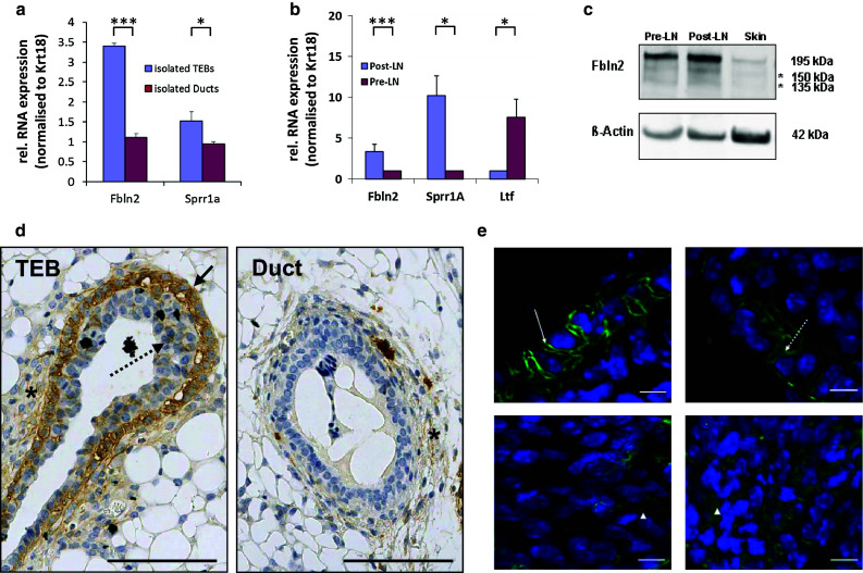
FBLN2 localises to cap cells of the TEB. Relative mean expression of Fbln2 mRNA in a isolated TEB and ducts and b in pubertal mammary tissue strips enriched for TEB (Post-LN) or ducts (Pre-LN) measured by qRT-PCR and normalised to Krt18. Mean expression of Fbln2 is significantly greater in TEB than ducts. The TEB marker Sprr1a and ductal marker Ltf were used as controls for TEB or ducts as described previously [19, 51]. Samples in a were pooled from 300 mice (1,145 TEBS and 1,111 ducts) as described in [19] and error bars denote technical variability. Error bars in b indicate 95 % confidence levels for at least three qRT-PCRs using at least three independent RNA extracts from different animals. *p value ≤0.05, ***p value ≤0.001. c Western Blot for FBLN2 protein (195 kDa) using rabbit polyclonal anti-FBLN2 antibody in the Pre-LN strips, Post-LN strips and skin (positive control). The image is representative of three independent experiments. Asterisks denote two additional, uncharacterised bands of a lower molecular weight (150, 135 kDa), most likely proteolytic cleavage products. β-actin was used as loading control. d Immunohistochemical staining for FBLN2 on FFPE-tissue sections of pubertal mouse mammary glands, showing strongest FBLN2 expression in the outer cap cell layer of TEB (black arrow). Some protein is also detectable between body cells (dotted arrows) and in the stroma around TEB (asterisk). Expression localises to the cell membranes, cytoplasm and extracellular spaces. Very low FBLN2 levels can be detected within the stroma around ductal epithelium (asterisk). Scale bars are 100 μm. e FBLN2 detected by confocal immunofluorescence microscopy on FFPE-tissue sections of pubertal 6 week old mice showing detailed FBLN2 localisation to cap cell (upper row) membranes (arrow) and cytoplasm (dashed arrow). Body cells (lower row) show FBLN2 staining in the cytoplasm or extracellular spaces (arrow heads). Scale bars are 10 μm. The images are representatives of six glands examined from three animals
