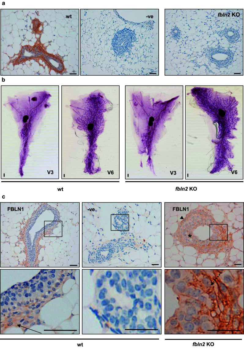
Fig. 8.

Fbln2 KO mice have no obvious mammary phenotype but express higher levels of FBLN1. a FBLN2 stained sections from six week mammary glands collected from wild type (wt) and Fbln2 KO mice (Fbln2 KO). No FBLN2 staining was detected in the no-primary antibody control (−ve) or in Fbln2 KO mouse mammary glands whereas prominent FBLN2 expression was seen in wt epithelium. Scale bars are 50 μm. 3 Fbln2 KO and 3 wt mice were examined. b Representative carmine alum stained wholemounts from 3 week and 6 week wt (left panel) and Fbln2 KO (right panel) mice. Scale bars are 1 mm. c Up-regulation of FBLN1 in the epithelium of Fbln2 KO pubertal mouse mammary glands. Mammary glands of pubertal 6 week wt (wt) and Fbln2 KO (Fbln2 KO) mice showing TEB epithelium stained with anti-FBLN1 (Fbln1) or no primary FBLN1 antibody (−ve). The lower row shows the corresponding magnified areas of each image. Weak FBLN1 staining in wt glands was contained in the stroma around TEB (arrow). In Fbln2 KO glands, however, FBLN1 was strongly detectable, localising to the basement membrane (arrow head), cap cells (dotted arrow) and cytoplasm and extracellular spaces between body cells (asterisk), in similar locations to those seen for FBLN2 in wt pubertal mouse mammary glands. Scale bars are 50 μm (wt) and 25 μm (KO). n = 3 animals
