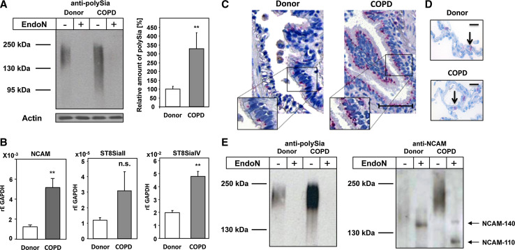
Fig. 4.
Expression and distribution of polySia-NCAM in lungs of COPD patients. a PolySia was analyzed in lung lysates of healthy donors and COPD patients by Western blotting. As specificity control, lysates were exposed to endoN prior to analysis. As loading control actin was used. Apparent molecular masses of standard proteins are indicated in kDa. Protein bands representing polySia were quantified by densitometry, and values are means of eight healthy donors and ten COPD patients each (100 % was set for healthy donors). b mRNA expression levels of NCAM, ST8SiaII and ST8SiaIV in lungs of donor and COPD patients were determined by quantitative real-time PCR; GAPDH was used as standard housekeeping gene. Values represent means of three donors and 11 COPD patients, respectively. The statistical evaluation was performed by Student’s t test (unequal variances, two-tailed). Significance levels are indicated by n.s. (not significant), p > 5 %, *p < 0.05, **p < 0.01, ***p < 0.001. c, d The distribution of polySia was visualized with anti-polySia mAb on paraffin embedded lung section from healthy donors and COPD patients; scale bar equals c 50 μm and d 25 μm. e Polysialylated NCAM were purified from lung lysates of donor and COPD patients and visualized by Western blotting using a mAb against polySia and an anti-NCAM (mAb 123C3) before and after polySia degradation by endoN

