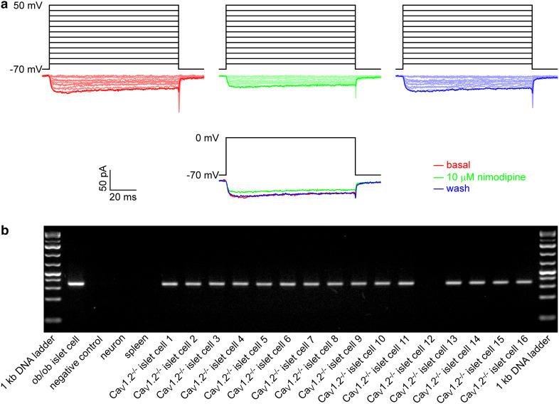
Fig. 1.
CaV1.3 channels are functionally expressed in a subgroup of mouse islet CaV1.2−/− β cells. a Examples of whole-cell CaV current traces evoked by a set of depolarizing voltage pulses (upper panel) and those generated by single voltage pulses (lower panel) in an islet cell from the β cell-specific CaV1.2−/− mouse before (red), during exposure to 10 µM nimodipine (green) and after washing treatment (blue). b RT-PCR analysis of cDNA obtained from single islet cells of the β cell-specific CaV1.2−/− mouse and from the positive control ob/ob islet cell and negative controls neuron, spleen and sterile ultrapure water with specific primers for insulin (344-bp amplicon)

