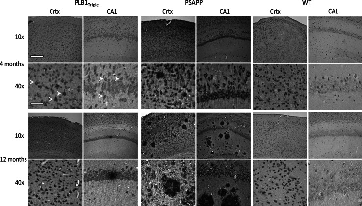
Fig. 9.
Amyloid pathology in PLB1Triple and PSAPP mice at 4 and 12 months old. PLB1Triple mice demonstrate intracellular amyloid in both the cortex (Crtx) and hippocampus (CA1), with infrequent extracellular plaque deposition and increasing amyloid reactivity with age. In comparison, PSAPP mice demonstrate a more aggressive expression of intracellular amyloid in both regions with a striking increase in plaque frequency with age. Age-matched wild types (WT) shown for comparison. Note the subtle intracellular staining for amyloid at 4 months in PLB1Triples (white arrows). Amyloid detected with 6E10 antibody (see methods for details). Scale bars represent 200 and 50 μm at 10× and 40× images, respectively

