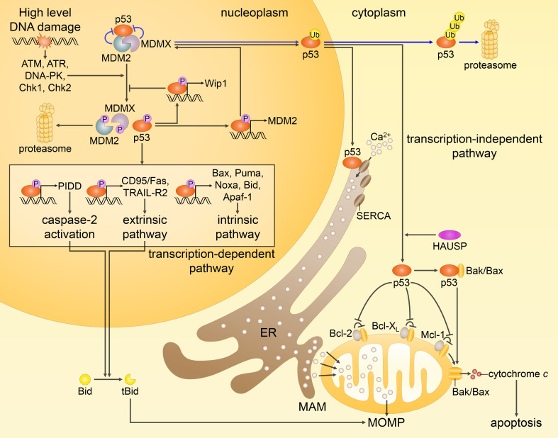
Fig. 3.
Different routes of p53-mediated apoptotic cell death. In response to extensive DNA damage, the interactions between p53, MDM2 and MDMX are disrupted by posttranslational modifications (only some phosphorylation events are shown), preventing inhibition and proteasomal degradation of p53 (p53 is normally kept at low levels by MDM2 and MDMX, as represented by the blue arrows). Activated p53 upregulates proapoptotic genes (as highlighted in the box), and represses antiapoptotic genes (not shown). As a result, p53 predominantly engages the intrinsic apoptotic pathway. In addition, p53 transactivates MDM2 and Wip1 phosphatase, providing the negative feedback loops that help to restrain p53 at the end of a stress response. Non-transcriptional activities of p53, as shown on the right, include translocation of its monoubiquitinated form to the mitochondria, where, after being deubiquitinated by HAUSP, it promotes Bak/Bax oligomerization through direct physical interaction with Bak/Bax or by binding of antiapoptotic proteins Bcl-2, Bcl-XL and Mcl-1. On the other hand, cytosolic p53 interacts with the sarco-ER Ca2+-ATPase (SERCA) pumps at the endoplasmic reticulum (ER) and mitochondrial-associated membranes (MAMs), potentiating Ca2+ influx followed by an enhanced transfer to the mitochondria. These transcription-independent functions of p53, along with the transcription-mediated events, prompt MOMP leading to the release of cytochrome c and subsequent apoptosis

