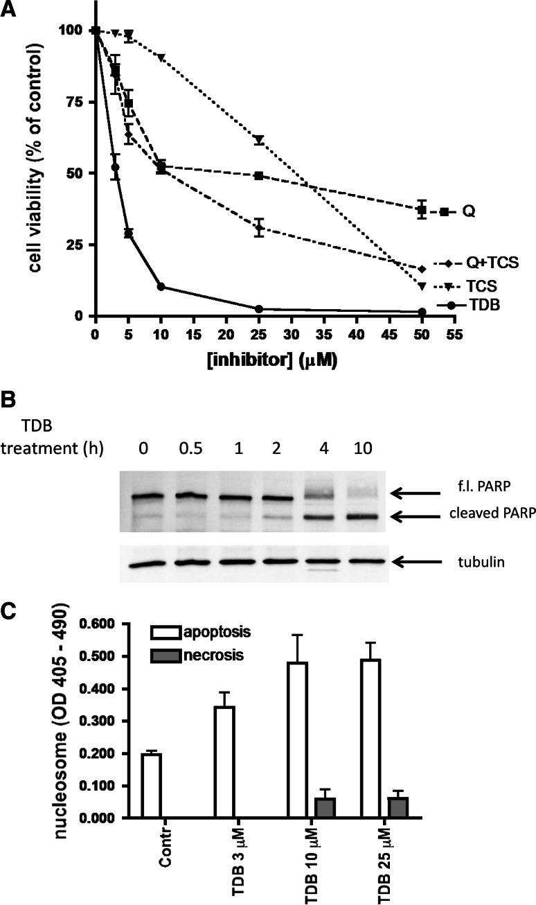
Fig. 6.
Effect of TDB on cell viability. a CEM cells were treated for 24 h with increasing concentrations of the CK2 inhibitor quinalizarin (Q), or the Pim-1 inhibitor TCS, or both (Q+TCS), or TDB. Cell viability was detected by the MTT method. Mean ± SEM values of four independent experiments are shown. b CEM cells were treated with 5 μM TDB for the indicated times, then apoptosis was assessed by analyzing PARP cleavage by Western blot of 10 μg of cell lysate proteins; tubulin was used as loading control. Representative results of at least three experiments are shown. c CEM cells were treated for 4 h with the indicated concentrations of TDB. The Cell Death Detection Elisa kit (Roche) was used for detecting nucleosome release in the cytosol (apoptosis) and in the extracellular medium (necrosis). Mean ± SEM of four experiments are shown

