Fig. 5.
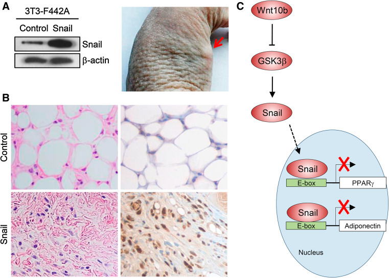
Overexpression of Snail inhibits adipogenesis in vivo. The 3T3-F442A cells were transfected with plasmids harboring Snail or backbone control plasmids, confirmed by immunoblots (a, left panel). After 2 days, preadipocytes (3 × 10⁷ cells) were harvested with trypsin and were injected subcutaneously into the flank area of athymic mice. After 5 weeks of implantation, a mass was developed at the site of injection. Red arrows indicate mass formation in the flank (a, right panel). Histology of the tissues isolated from athymic mice xenografts (magnification, ×400). The dissected mass was fixed in formalin, followed by staining with hematoxylin and eosin (H&E) (b, left column). Immunohistochemistry with anti-Snail antibody was also performed in the background of hematoxylin (b, right column). The left upper figure shows normal adipogenesis in 3T3-F442A cells of the control group, whereas the left bottom figure indicates that Snail-overexpressing cells were not differentiated into adipocytes in vivo. Immunohistochemistry shows Snail-positive brown nuclei in the tissue of mice injected with Snail-overexpressing cells (right bottom), while differentiated adipocytes in control mice have no expression of Snail. Figures are representative of four tissues. c Schematic illustration showing the putative mechanism by which Snail inhibits adipocyte differentiation

