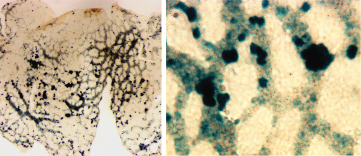
Fig. 1.
Runx1high clusters (β-galactosidase staining) in the E8.5–E9.0 yolk sac of Runx1-LacZ conceptuses [59], at lower magnification (left panel) and at higher magnification (right panel). The vitelline vascular plexus is filled with primitive erythroblasts that downregulate Runx1 at the time when their progenitors disappear. Note that even though all clusters are associated with nascent vasculature, some of them protrude into the avascular area. This is consistent with the model of separate emergence of blood and endothelial cells in the yolk sac

