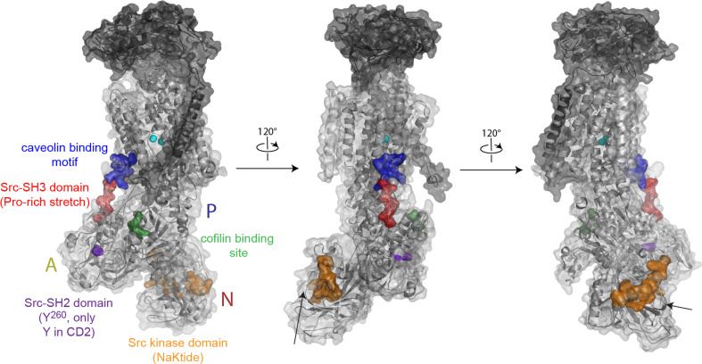
Fig. 3.
Potential binding sites located on the cytoplasmatic domains of Na+,K+-ATPase. For Na+,K+-ATPase αβγ-complex, an illustration and transparent surface are shown. The orientation of the left image is as in Fig. 2b. The A-, N-, and P-domains are indicated. Possible binding sites are shown by colored surface presentations. The NaKtide fragment is mainly located in the core of the N-domain (black arrows in middle and right figure) and hence are only partially accessible. Rb+ is shown in cyan spheres

