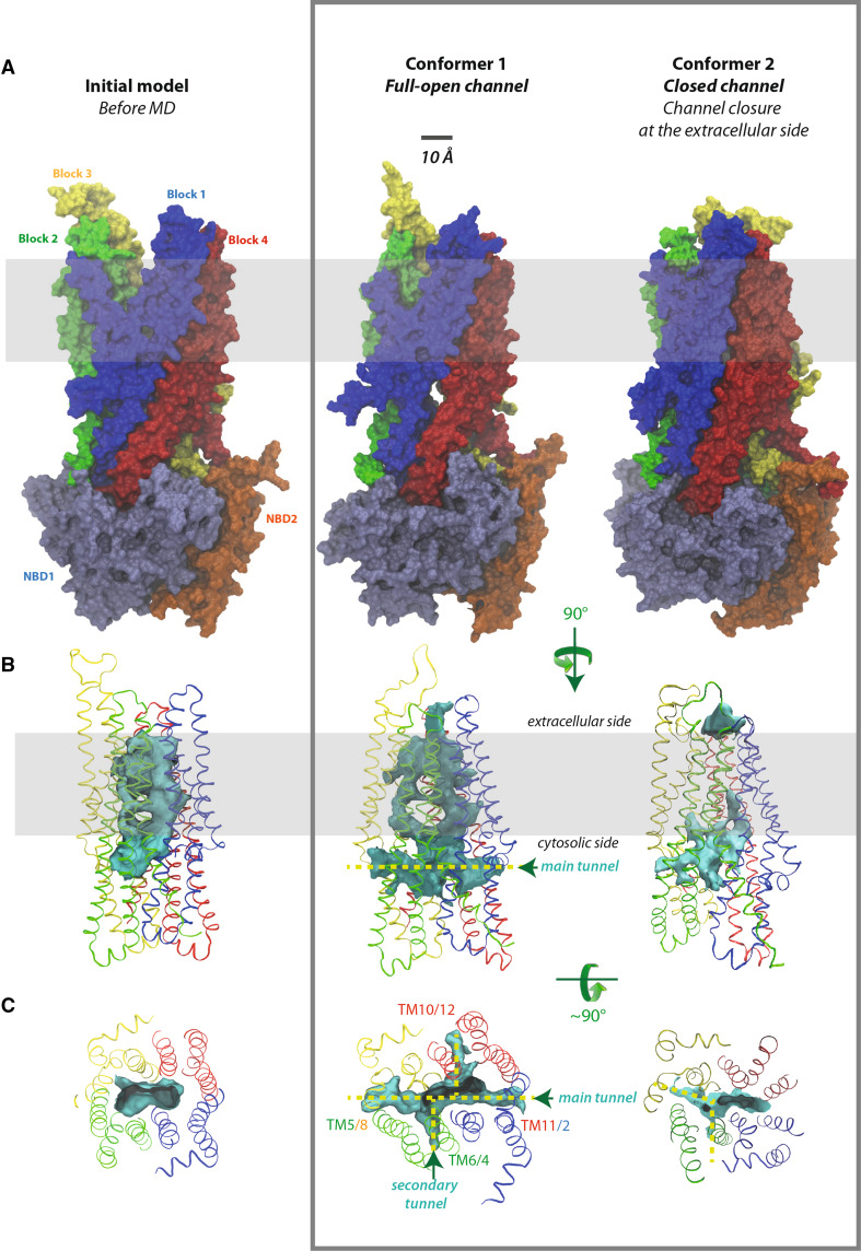
Fig. 2.
The channel and its cytosolic access routes—global views. A Solid representation of the CFTR MSD:NBD assembly of the initial model, before MD (at left), conformer 1 (at the center, full-open channel) and conformer 2 (at right, closed channel). The main lateral tunnel (~47 Å long) is visible below the membrane, through its TM11/TM2 entrance in conformer 1. The position of the lipid bilayer is symbolized in gray. B Ribbon representations of the MSD assembly (four three-helix blocks) of the three same models, encircling the water-filled cavity constituting the ion channel (in solid, colored in blue green). The transition between the initial model (at left) and conformer 1 (at the center) gives rise to a well-defined channel and, importantly, to cytoplasmic lateral tunnels allowing access from the cytosol and merging with the channel. Note that the channels are arbitrarily cut at the extracellular sides, as they reach there the solvent. C Orthogonal view of the cytosolic end of the channel and of the lateral tunnels. These are labeled according to the TM helices participating in their formation. There are only two cytosolic entrances in conformer 2

