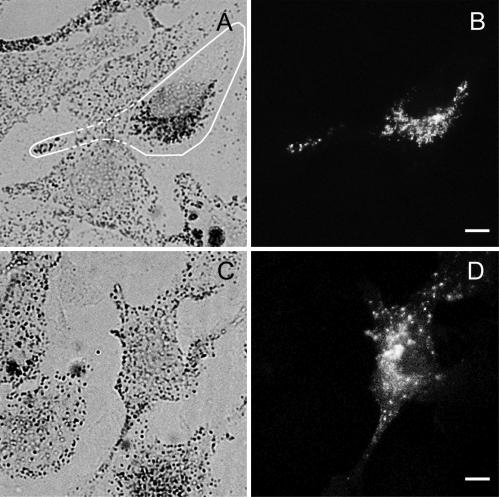
Figure 4.
MC STΔD, but not MC STΔF, colocalizes with melanosomes and generates a dominant negative phenotype in wild-type melanocytes. Shown is the distribution of melanosomes (A and C) and GFP fluorescence (B and D) in melan-a melanocytes transfected with either GFP-MC STΔD (A and B) or GFP-MC STΔF (C and D). The edges of the transfected cell in A are marked with a white line. Bars, 6 μm.

