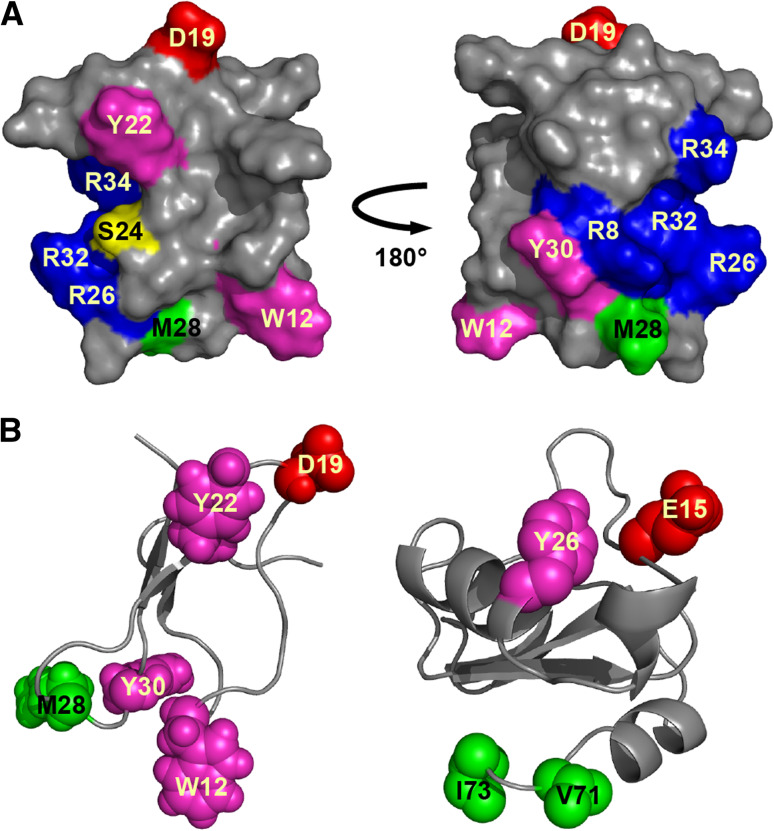
Fig. 4.
Structure of site-4 NaV channel toxins. a Surface representation of the spider toxin δ-amaurobitoxin-Pl1b (PDB 1V91) showing key pharmacophore residues. Adapted from [45]. b Ribbon representation of δ-amaurobitoxin-Pl1b (left) and the scorpion toxin Bj-xtrIT (PDB 1BCG, right) showing the similar spatial positioning of key functional residues. In each panel, the chemical nature of amino acid residues is color coded as follows: aromatic, magenta; aliphatic, green; basic, blue; acidic, red; polar but uncharged, yellow

