Fig. 1.
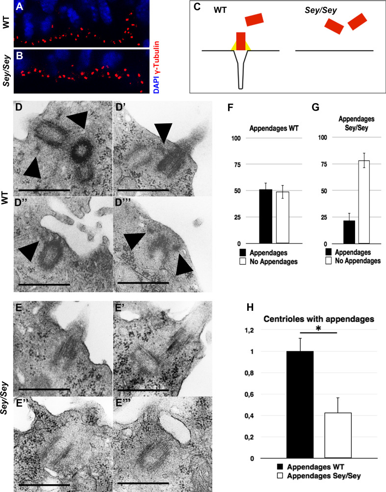
Defect of centrosome maturation in Pax6-deficient cortex. a, b IHC with the centrosome marker γ-Tubulin (in red) in WT (a) and Pax6/Small eye (Sey/Sey) (b) E15.5 cortex reveals a disturbed localization of centrosomes in the mutant cortex. Cell nuclei are counterstained with DAPI (blue). c Schematic presentation to explain how a structural defect of the mother centriole appendages could lead to mis-localization of the centrosomes. d–e′′′ Electron microscopy micrographs illustrating the structural defect of the mother centrioles in Sey/Sey (e–e′′′) compared with WT (d–d′′′) control cortex. Arrowheads point to subdistal appendages easily detectable only in WT cortex. f–g Statistical analysis of the centriole number at the ventricular surface of WT embryo showing that half of the centrioles (51.26 ± 6.21 %) contains appendages, thus identifying them as mother centrioles, and 48.74 % (±6.21) as daughter centrioles (without appendages). Note that in the Sey/Sey cortex only 21.8 % (±7.13) of centrioles contain appendages and 78.2 % (±7.13) miss these appendages (n = 3) (bars 0.5 µm). h Relative to the control, the Pax6 deficient cortex shows a strong reduction in the number of matured centrioles containing appendages at the ventricular surface (WT: 1 ± 0.12); Sey/Sey: 0.43 ± 0.14); *≤0.05; p = 0.012)

