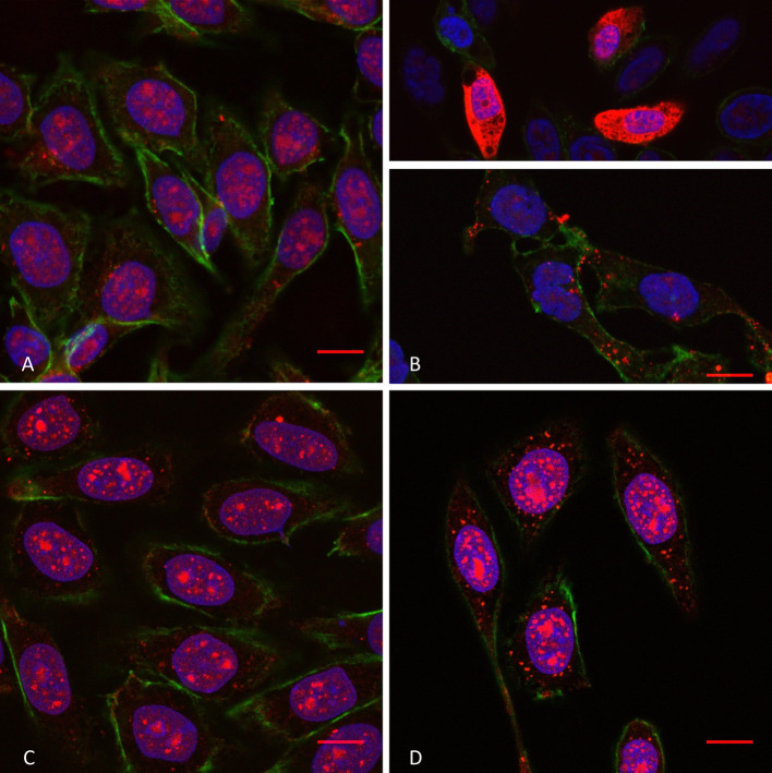
Fig. 3.
Confocal microscopy of penetratin internalisation (10 µM extracellular concentration) in WT cells in the absence (a) and the presence (b) of SMase, and in GAGneg cells in the absence (c) and the presence (d) of SMase. The peptide is labelled in red (streptavidin–TRITC), the actin cytoskeleton protein in green (phalloidin–FITC) and the cell nuclei in blue (DAPI). Scale bar corresponds to 10 µm. Unmerged pictures are shown in Supplementary Fig. 3

