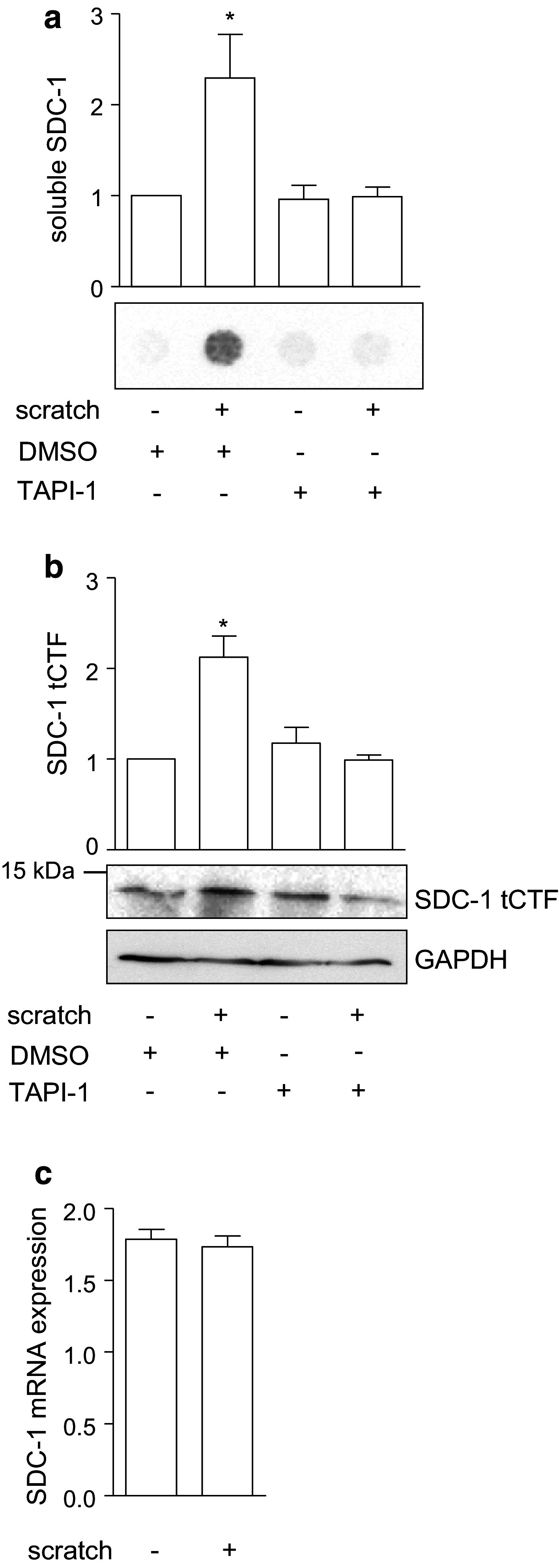
Fig. 2.

Association of syndecan-1 shedding and wound closure in A549 cells. a–c A confluent monolayer of A549 cells was wounded by a defined scratch or left untreated. The cells were incubated for 24 h with metalloproteinase inhibitor TAPI-1 (10 µM) or DMSO (0.1 %) as control. Subsequently, the supernatants were analyzed for the presence of soluble SDC-1 by dot blotting using an antibody against the ectodomain of SDC-1 (a) and cell lysates were subjected to Western blotting using an antibody against SDC-1 tCTF (b). Signals were quantified by densitometry and expressed in relation to the DMSO treated control (a–b). The expression of full length SDC-1 mRNA was controlled by quantitative PCR (c). Data are shown as representative experiments and means + SD calculated from three independent experiments. Statistically significant differences compared to the control without scratch induction are indicated by asterisks (p < 0.05)
