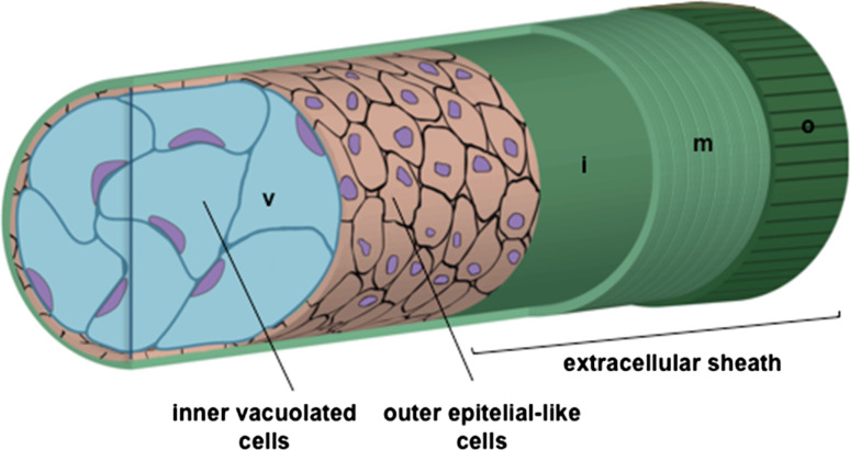
Fig. 1.
Structure of the zebrafish notochord and its surrounding sheaths. This cartoon illustrates notochord structure of inner vacuolated cells surrounded by an epithelial-like sheath of cells. Around these cells, a peri-notochordal BM is present. This extracellular sheath is composed of three parts: the inner (i) basal lamina, surrounded by a medial (m) layer of collagen fibrils that run parallel to the notochord, and an outer (o) granular layer of loosely organized matrix that is mostly oriented in a perpendicular manner to the notochord. The peri-notochordal sheath counteracts the pressure generated from vacuoles (v), providing the rigidity and the correct stiffness of this structure

