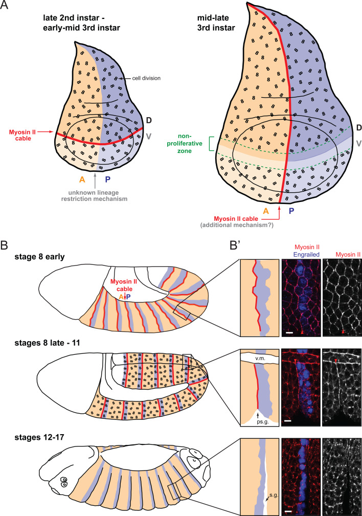
Fig. 2a–b′.
Actomyosin cables at compartmental boundaries. a In the Drosophila wing disc, an actomyosin cable is present at the D/V boundary from late second instar to mid third instar (red line). At that time, the boundary is challenged by cell division (represented by “8” symbols). The D/V actomyosin cable is dismantled in mid third instar larvae (around 96 h AEL) when a nonproliferating zone forms that straddles the boundary (green dashed lines). At the same time, another actomyosin cable forms at the A/P boundary. The mechanism that implements cell sorting at the A/P boundary at earlier stages is currently unknown. b, b′ In the embryo, parasegmental actomyosin cables are detectable by early stage 8, before cell division resumes in the epidermis at stage 8. They are dismantled by stage 12, once cell division ceases in the epidermis. When parasegmental actomyosin cables form at early stage 8, the boundary is not straight yet, but it straightens by stage 9 (top and middle panels in b′). This is consistent with the in silico modelling of local increase in tension between two cell populations performed by Landsberg and coworkers [49]. Also, consistent with increased tension at the parasegmental boundary, a “parasegmental” groove (ps.g.) forms at stage 10 that correlates spatially and temporally with parasegmental actomyosin cables (middle panel in b′). Scale bar in b′: 5 μm. v.m. Ventral midline, s.g. segmental groove. b′ is adapted by permission from Macmillan Publishers Ltd: Nature Cell Biology [50], copyright 2010

