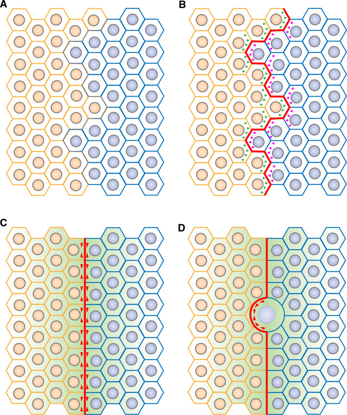
Fig. 4a–d.
A model for cell sorting at compartmental boundaries: role of contractile actomyosin barriers. a Patterning of fields of cells (such as segmentation in the Drosophila embryo or in the vertebrate hindbrain) leads to the apposition of two compartments. b Bidirectional signalling across the boundary (green and pink arrows) induces actomyosin cabling (red) at the compartmental boundary interface. c Cortical tension generated by the actomyosin cable (red arrowheads) at the level of adherens junctions straightens the compartmental boundary, leading to a segregation of cells based on their identity. A secondary organizer (green gradient) then triggers differentiation of the surrounding tissue. d Next, actomyosin cable-dependent cortical tensile forces correct local cell invasion, in particular following cell division, thereby maintaining a straight boundary and stable compartmentalization

