Fig. 2.
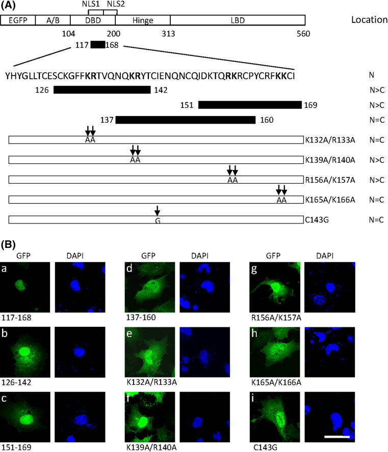
Detection of key basic residues in NLS1 for nuclear import. a Sequence of the putative NLS1 is shown. The full-fragment (residues 117–168) and three segments within this region (residues 126–142, 137–160 and 151–169) were fused to EGFP. The basic amino acids lysine and arginine (in boldface) in the 117–168 region were substituted by alanines as indicated by arrows. The subcellular localization of LRH-1 mutants is summarized on the right (N nuclear, C cytoplasmic). b Wild-type and mutant forms were transfected into COS-7 cells. After 24 h, cells were fixed and the nuclei counterstained with DAPI. Fusion proteins were visualized by confocal microscopy. Scale bar 25 μm

