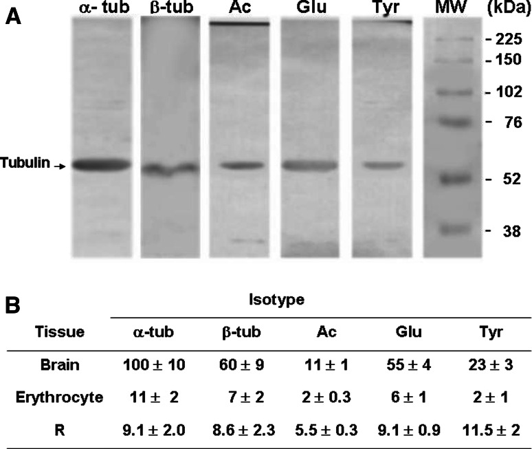
Fig. 1.
Presence of tubulin in human erythrocytes. Sedimented erythrocytes prepared from 2 ml blood were lysed by resuspension in 3 ml lysis buffer containing 1% Triton X-100. a Aliquots (250 μg protein) of lysate were analyzed by SDS-PAGE and immunoblotted using antibodies specific to total α-tubulin (α-tub), β-tubulin (β-tub), tyrosinated tubulin (Tyr-tub), detyrosinated tubulin (Glu-tub) and acetylated tubulin (Ac-tub). b Tubulin bands were quantified (optical density in arbitrary units) using the Scion Image program, with optical density of the total α-tubulin band defined as 100%. Values are expressed as mean ± SD from three independent experiments. Homogenate samples prepared from 30-day-old rat brain were processed in parallel for comparative purposes. The brain sample is not shown in a

