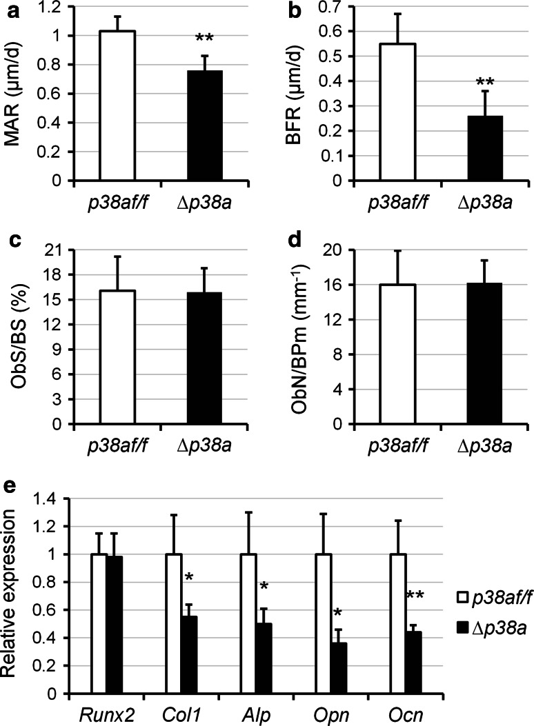
Fig. 4.
Osteoblast-specific disruption of p38a decreases bone formation in mice. a–d Quantitative histomorphometric measurements were performed on the spongiosa at distal femurs of 12-week-old female p38a f/f and ∆p38a mice (n = 6 per group). a MAR mineral apposition rate; b BFR bone formation rate; c ObS/BS osteoblast surface/bone surface; d ObN/BPm osteoblast number/bone perimeter. e Real-time PCR analyses of osteoblast marker gene expression in femurs of 12-week-old female p38a f/f and ∆p38a mice (n = 6 per group). *p < 0.05, **p ≤ 0.01

