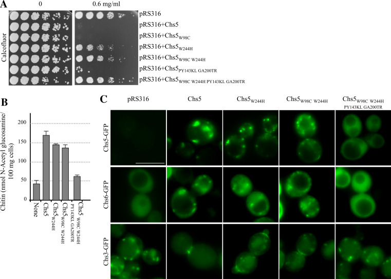
Fig. 4.
Conserved residues in the FN3 and BRCT domains are relevant for Chs5p localization and functionality. a A total of 3 × 10⁴ cells and serial 1:4 dilutions from a chs5Δ strain bearing the indicated plasmids were spotted onto buffered SD-URA plates supplemented with the indicated amounts of Calcofluor and incubated for 2 days at 32ºC. The Chs5 proteins were untagged. b Amount of chitin in a chs5Δ strain bearing centromeric plasmids producing the indicated untagged Chs5 proteins. c Localization of the indicated Chs5 proteins fused to the GFP in chs5Δ cells (upper row panels) or localization of GFP-fused Chs6p (middle row panels) and GFP-fused Chs3p (lower row panels) in chs5Δ cells expressing the indicated untagged Chs5 proteins from centromeric plasmids. Cells in the micrographs show the most representative distribution of the GFP-fused proteins in each strain. Bar 10 μm

