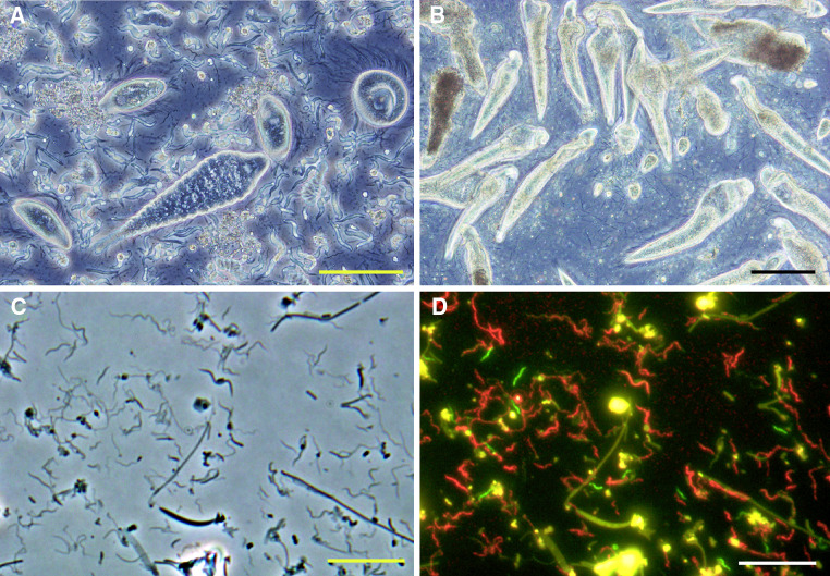
Fig. 2.
Microbiota in the hindgut of termites. a Phase-contrast image of the microbiota in the gut of the lower termite Reticulitermes speratus. b Phase-contrast image of the microbiota in the gut of the lower termite Coptotermes formosanus. c Phase-contrast image of the microbiota in the wood-feeding higher termite Nasutitermes takasagoensis. d FISH (fluorescent in situ hybridization) image of c. Texas-red signals: treponemes; 6FAM (green) signals: a novel group (FibS2) of the phylum Fibrobacteres [72]. Amorphous yellow colors were autofluorescence emitted from wood particles. Bars = 100 μm in a and b and 10 μm in c and d

